38 fluid mosaic model diagram
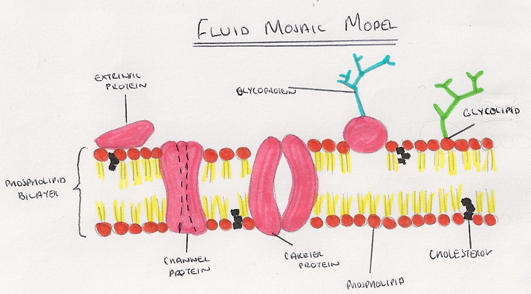
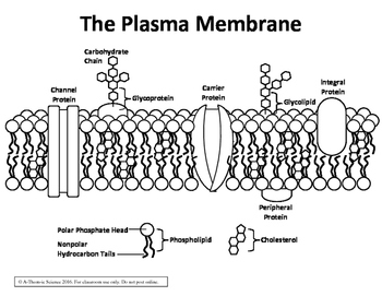
Draw a labelled diagram of the fluid mosaic model of the plasma membrane. The cell membrane is a multifaceted membrane that envelopes a cell's cytoplasm. The interior of a cell in between the plasma membrane and the nucleus is filled with a semifluid product called cytoplasm. A channel protein is an example of an. The specialized structure of the cell membrane described by the fluid mosaic model allows the cell to be selectively permeable, with dynamic homeostasis being maintained by constant movement of molecules across the membrane. Double check that your analogies work together just like the organelles work together in a cell.
fluid mosaic model. model that describes the arrangement and movement of the molecules that make up a cell membrane. Sets found in the same folder. Membrane Structure.

Fluid mosaic model diagram
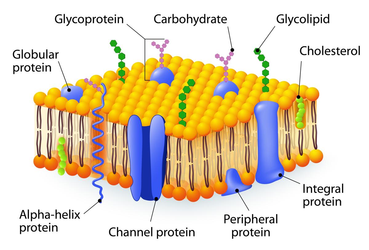
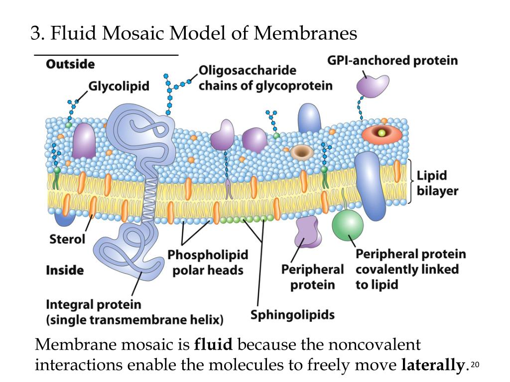
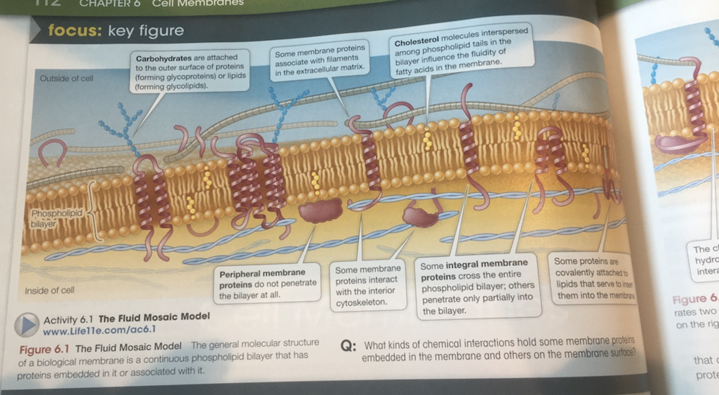
The Fluid Mosaic Model The fluid mosaic model is the current accepted model for the structure of the cell membrane, In this model the membrane is fluid as the phospholipids are constantly moving. The other components of the membrane (such as the proteins) are scattered throughout the bilayer like a mosaic. This student diagram shows the arrangement of molecules in the fluid mosaic model of cell membranes. Which of the following correctly identifies the labels A to D? A = integral protein, B = cholesterol, C = phospholipid, D = peripheral protein. Fluid mosaic model was proposed by Singer and Nicolson in 1972. According to this model, the quasi-fluid nature of lipids enables lateral movement of proteins within the overall bilayer. This ability to move within the membrane is measured as its fluidity.
Fluid mosaic model diagram. According to the fluid mosaic model, the membrane is an ever-changing structure in which the mosaic protein floats on the lipid bilayer acting as a fluid. Proteins in this model do not form a continuous layer covering both sides of the membrane as proposed by Daniel and Davson model. The plasma membrane may be known as a fluid mosaic model where the membrane is a fluid structure with various proteins embedded in or attached to the bilayer of phospholipids. The plasma membrane possesses hydrophilic tails and hydrophobic tails, which may be referred to as amphiphilic. Danielli and Davson proposed a model whereby two layers of protein flanked a central phospholipid bilayer. The model was also described as a 'lipo-protein sandwich', as the lipid layer was sandwiched between two protein layers. The Davson-Danielli model predominated until Singer and Nicolson advanced the fluid mosaic model in 1972. The currently accepted model of the membrane is the Fluid Mosaic Model described by Singer and Nicholson. One of the foremost elaborate responsibilities that healthiness authorities face throughout their interplay with patients helps them recognise the issues and a way to encourage them concerning the prognosis and treatment available.
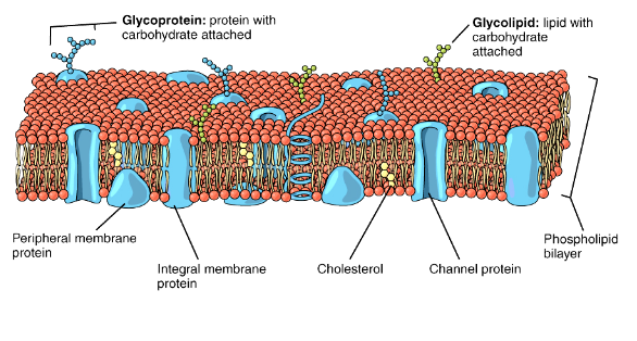
Fluid mosaic model MCQs. Hello students, our next topic of MCQ is 'Fluid mosaic model'. Cell membranes are made up of three main components that are phospholipids, cholesterol and proteins. Scientists viewed this cell wall under microscope and given name that is fluid mosaic model. Fluid Mosaic Model of Cell Membrane. The cell membrane consists of a phospholipid bilayer oriented with the hydrophobic fatty acid tails facing the middle of the bilayer, and the hydrophilic polar heads facing interior and exterior surfaces. Proteins, glycoproteins, and glycolipids are embedded in the membrane, and cholesterol is inserted into the lipophilic interior. The fluid mosaic model is a model that scientists use to describe the structure of the plasma membrane. The plasma membrane is fluid, meaning that the components drift around freely in a lateral way. Make sure you can draw and label all the above structures on a diagram of the fluid mosaic model of cell membranes. List the diagrams in order from first to last in the cell cycle. Colorful animations make these flash games as fun as it is educational. The study of cells is called cell biology cellular biology or cytology.
Based on the structure of the plasma membrane, it is regarded as the fluid mosaic model. According to the fluid mosaic model, the plasma membranes are subcellular structures, made of a lipid bilayer in which the protein molecules are embedded. Also refer to the Difference Between Cell Membrane and Plasma Membrane Draw and label a simple line diagram of a cell membrane. Cell membrane functions role structure video lesson. 50 Cell Membrane Coloring Worksheet Answer Key Vz9b di 2020 Draw a labelled diagram of the fluid mosaic model of the plasma membrane. How to draw a cell membrane. Structure and composition of the cell membrane. Proteins […] Mosaic – the phospholipid bilayer is embedded with proteins, resulting in a mosaic of components. Structure of the Plasma Membrane (Fluid-Mosaic) Components of the Plasma Membrane. Phospholipids – Form a bilayer with phosphate heads facing outwards and fatty acid tails facing inwards. Cholesterol – Found in animal cell membranes and functions to improve stability and reduce fluidity. Fluid Mosaic Model Student Worksheet Answers - To help autistic youngsters reinforce their language abilities and also comprehend a range of subjects, an option of checking out understanding worksheets are offered. Pupils with autism, in addition to their instructors, would substantially benefit from these devices.
Apr 05, 2021 · The Fluid Mosaic Model of Biomembranes. The fluid mosaic model describes the structure of the plasma membrane as a mosaic of components —including phospholipids, cholesterol, proteins, and carbohydrates—that gives the membrane a fluid character. Membranes are impermeable to most polar or charged solutes, but permeable to nonpolar compounds ...
Fluid Mosaic Model Diagram The Fluid Mosaic Model Of The Cell Plasma Membrane F Cell Membrane Coloring Worksheet Cell Membrane Structure Fluid Mosaic Model . Ocr Biology Revision Part 3 Cell Membranes Biology Revision Cell Membrane Biology .
3. The most accepted structure is called the 'fluid mosaic' model, proposed by Singer and Nicolson. 4. Being selectively permeable regulates the movement of molecules. Nucleus: 1. The brain of the cell. This has a double membrane surrounding the nucleoplasm. 2.
The Fluid Mosaic Model states that membranes are composed of a Phospholipid Bilayer with various protein molecules floating around within it. A bacteria diagram basically enables us to learn more approximately this unmarried cell organisms that have neither membrane-bounded nucleolus or organelles like mitochondria and chloroplasts.
Fluid Mosaic Model Diagram The Fluid Mosaic Model Of The Cell Plasma Membrane For Du Cell Membrane Coloring Worksheet Cell Membrane Structure Cell Membrane . Medical Education Unit Mbbs 1st Year Biochemistry Paper2 2016 Of The Jaba Biochemistry Medical Education University Exam .
Time: 1h Students will learn how to draw and label a diagram of the fluid mosaic model of membranes. Using a webcast and revision flashcards the teacher has more freedom to assist students individually. Falsification of membrane structures. Time: 1h The accepted model of membrane structure today is the fluid-mosaic model but this has not always ...
The photograph or diagram of metaphasicchromosome arranged in homologous pair according to their length and thickness, position of centromere is called ... (d) None of these. Answer. Answer: (c) Idiogram. Question 14. The structure of plasma membrane fluid mosaic model is proposed by (a) Gram (b) Singer and Nicolson (c) Schwann and Schleiden (d ...
Describe the fluid mosaic model of membrane. Solution: The characteristic features of fluid mosaic model • This model was proposed by Singer and Nicholson. • According to this model, there is a central bilipid layer (of phospholipids) with their polar head group toward the outside and the non-polar tails pointing inwards.
Dec 04, 2021 · The fluid mosaic model describes the structure of the plasma membrane as a mosaic of components —including phospholipids, cholesterol, proteins, and carbohydrates—that gives the membrane a fluid character. Membranes are impermeable to most polar or charged solutes, but permeable to nonpolar compounds ... Start studying Fluid Mosaic Model Diagram. Learn vocabulary, terms, and more with flashcards, games, and other study tools.
Fluid mosaic model is the most accurate model that explains the structure of the cell membrane. Their proposal was the fluid mosaic model, which is the dominant model now. Both phospholipid molecules and embedded proteins are able to diffuse rapidly and laterally in the membrane.
Explain the fluid mosaic model of the plasma membrane? Differentiate between a prokaryotic and eukaryotic cell. Frequently Asked Question: FAQs. Ques: What is the NCERT Class 11 Chapter 8 CBSE Biology about? Ans: The name of the Class 11 Chapter 8 CBSE Biology Chapter 8 is Cell- The Unit of Life. This chapter helps the students in understanding ...
Fluid mosaic model of cell membrane state that it has lipid bilayer with (a) proteins on the outer surface only. (b) Some proteins embedded and some on the surface. (c) Proteins on both the surfaces. (d) Proteins embedded and some on the surface. Answer. Answer: (b) Some proteins embedded and some on the surface.
Membranes are described as a fluid lipid bilayer with floating proteins and carbohydrates in the Fluid Mosaic Model. Glycolipids and glycoproteins are chemically linked to membrane carbohydrates. Certain membrane carbohydrates, or else, are part of proteoglycans that inject their amino acid chain between lipid fatty acids.
Phospholipid Bilayer Structure. The phospholipid bilayer is made of two layers of phospholipids. Each phospholipid has a hydrophilic head and a hydrophobic tail.
Fluid mosaic model was proposed by Singer and Nicolson in 1972. According to this model, the quasi-fluid nature of lipids enables lateral movement of proteins within the overall bilayer. This ability to move within the membrane is measured as its fluidity.
This student diagram shows the arrangement of molecules in the fluid mosaic model of cell membranes. Which of the following correctly identifies the labels A to D? A = integral protein, B = cholesterol, C = phospholipid, D = peripheral protein.
The Fluid Mosaic Model The fluid mosaic model is the current accepted model for the structure of the cell membrane, In this model the membrane is fluid as the phospholipids are constantly moving. The other components of the membrane (such as the proteins) are scattered throughout the bilayer like a mosaic.









0 Response to "38 fluid mosaic model diagram"
Post a Comment