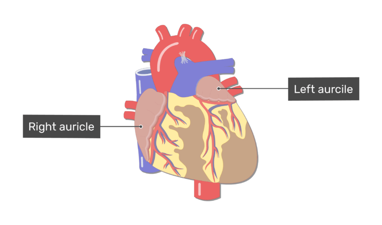
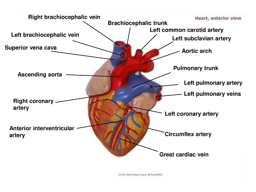
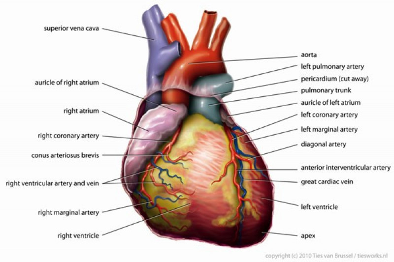
42 in the diagram, where is the left auricle of the left atrium?
A&P 2 - Cardiovascular System (heart, veins, & arteries) Identify the highlighted structures. Identify the highlighted structure.
Auricle of right atrium Right coronary artery (in atrioventricular sulcus) Right ventricle Inferior vena cava (a) Anterior view of the external heart C' 2019 Pearson Education. Aort'c arch Ligamentum arteriosum Left pulmonary artery Left pulmonary ve ns Auricle of left atrium Circumflex artery Left coronary artery (in atrioventricular sulcus)
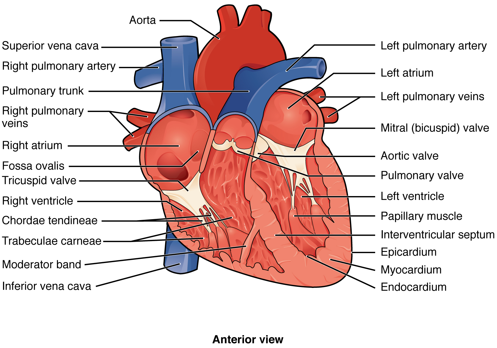
right atrium. left atrium. right auricle. left auricle. right ventricle. left ventricle. chordae tendineae. papillary muscles. interatrial septum. fossa ovalis. interventricular septum. tricuspid valve (right AV valve) Bicuspid Valve (Left AV Valve) pulmonary semilunar valve. aortic semilunar valve.

In the diagram, where is the left auricle of the left atrium?
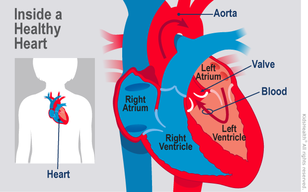
Now the impure deoxygenated blood is purified in the lungs after which this oxygenated blood is transported to the left auricle or atrium of the heart. This circulation of blood from the right ventricle to the left atrium of the heart is termed pulmonary circulation.
Left and right atrium Left and right ventricle Anterior and posterior interventricular sulcus Interventricular Septum Aorta Pulmonary trunk Pulmonary arteries (right and left) - Know them in a diagram only Pulmonary veins (2 right and 2 left) - Know them in a diagram only Superior and Inferior vena cava Tricuspid and Bicuspid (Mitral) valves
Left atrium: Receives oxygenated blood from the lungs via the pulmonary veins (4 ostia). Characteristics - Thicker wall; left auricle (contains pectinate muscles) Landmarks - T5 - T8 (supine), T6 - T9 (erect) Right atrium: Receives deoxygenated blood from the systemic circulation via the superior and inferior vena cava
In the diagram, where is the left auricle of the left atrium?.
Two pulmonary veins come from each lung and pass O 2 -rich blood to left atrium. Blood enters left ventricle from the left atrium. The left ventricle pumps blood into the aorta which in turn branches and delivers blood to the major body regions and organs. So, the correct sequence is C.
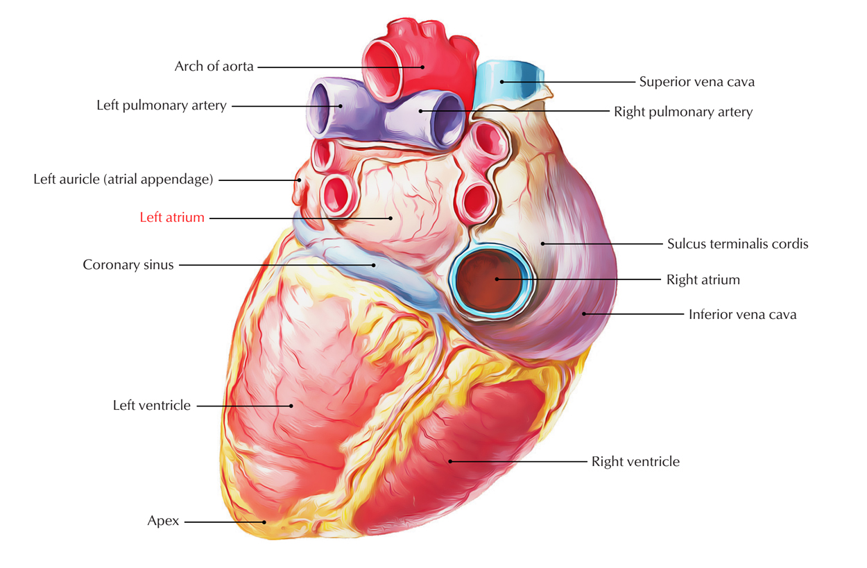
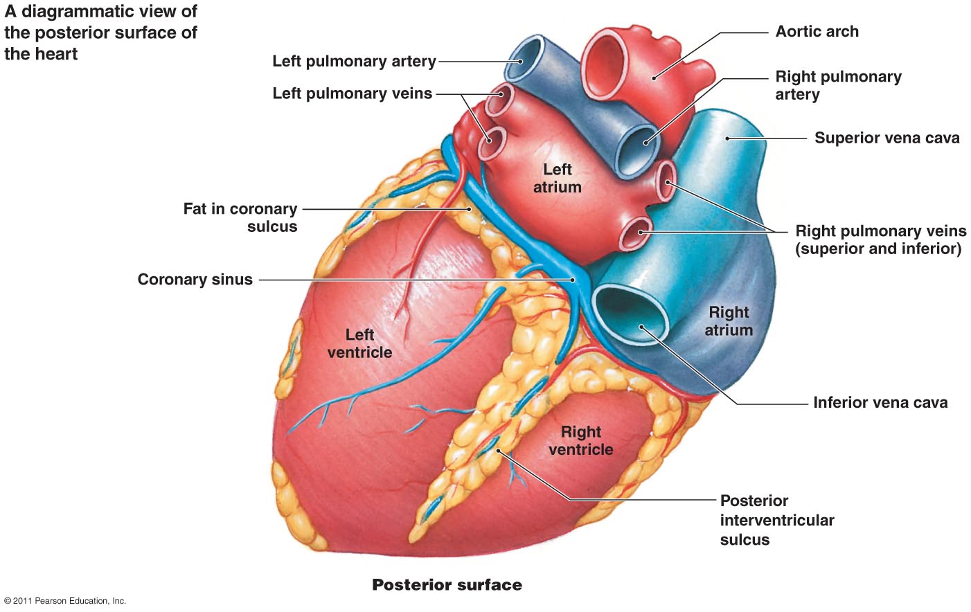
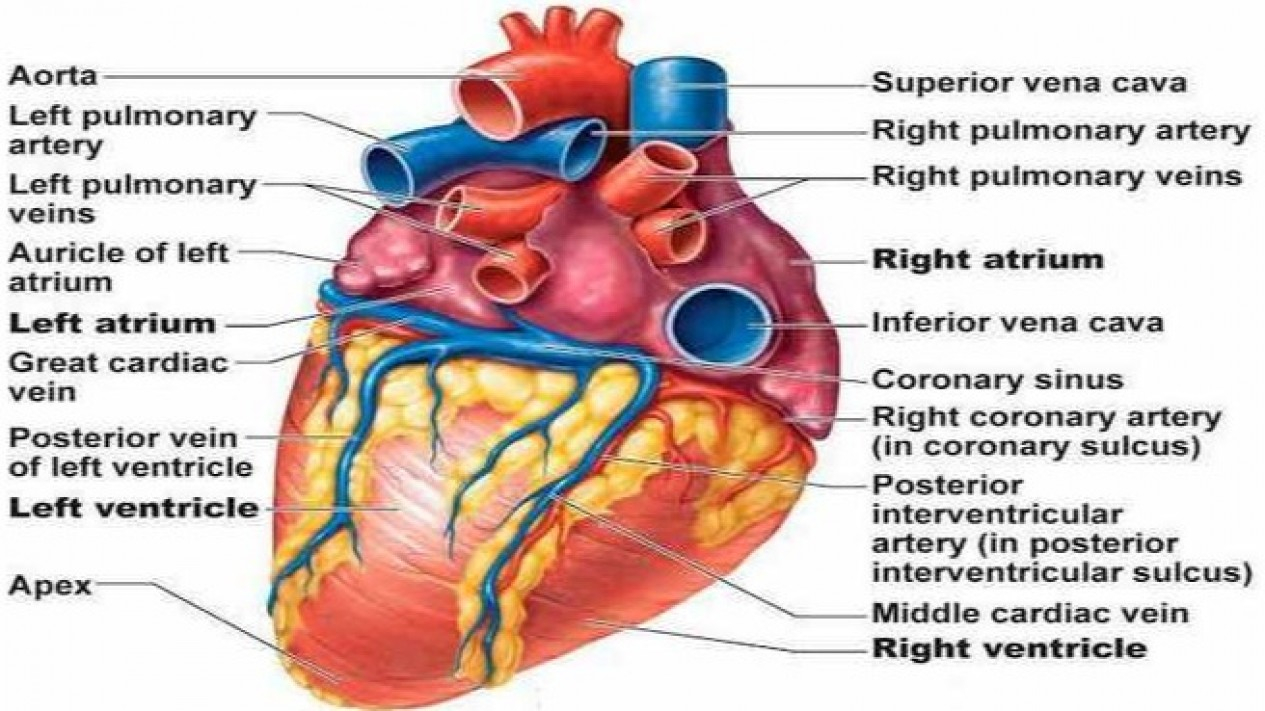
You may also find posterior interventricular sulcus, middle cardiac vein, right ventricle, apex, apex of heart, left ventricle, posterior vein of left ventricle, great cardiac vein as well. This picture also contains other parts such as left atrium, auricle of left atrium, posterior surface view of heart, left pulmonary vein, left pulmonary ...
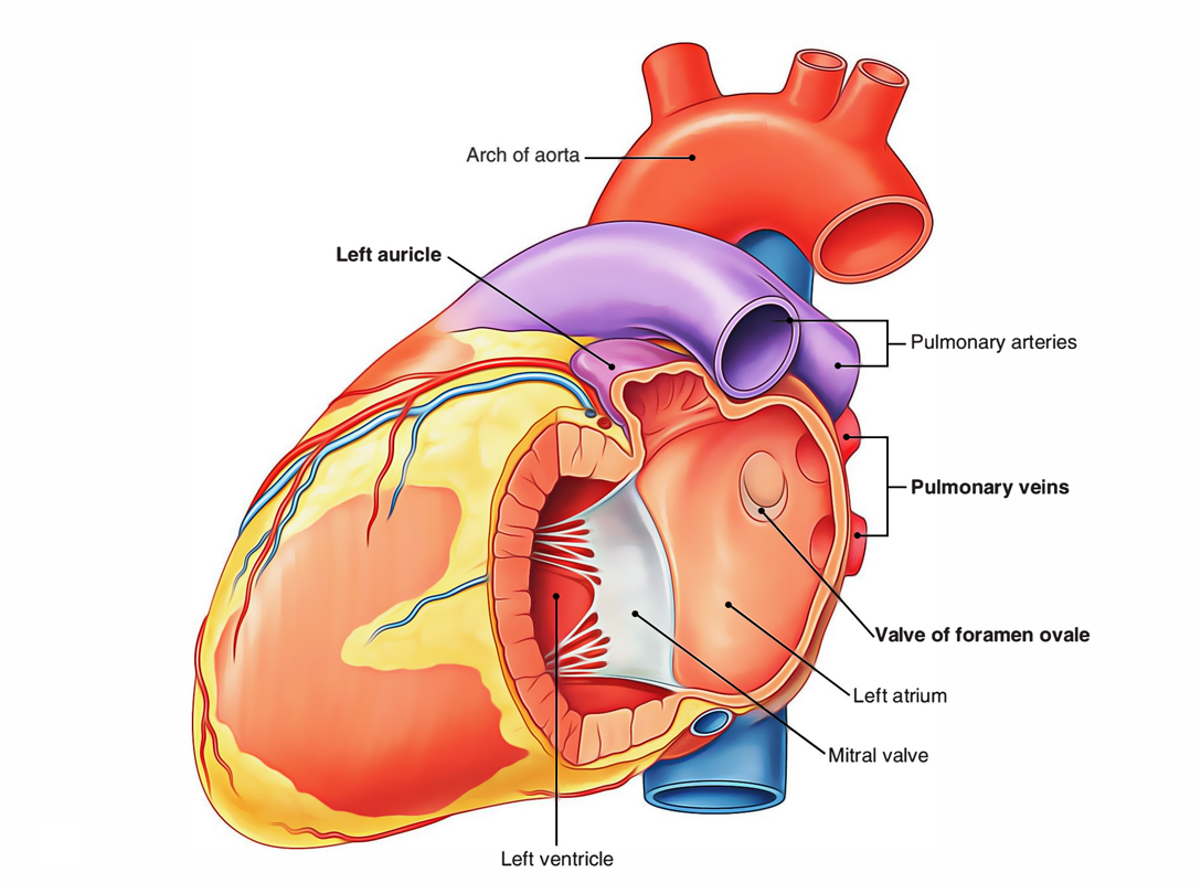
Left Atrium. The left atrium receives oxygenated blood from the four pulmonary veins, and pumps it through the left atrioventricular orifice (guarded by the mitral valve) into the left ventricle. In the anatomical position, the left atrium forms the posterior border (base) of the heart. The left auricle extends from the superior aspect of the chamber, overlapping the root of the pulmonary trunk.
The increase in effective pulmonary blood flow may be estimated by the left atrial to aortic ratio. The LA:Ao uses the relatively fixed diameter of the aorta to assess the degree of left atrial volume loading. This ratio correlates significantly with increased pulmonary flow attributable to excessive transductal flow; optimal threshold ratio ...
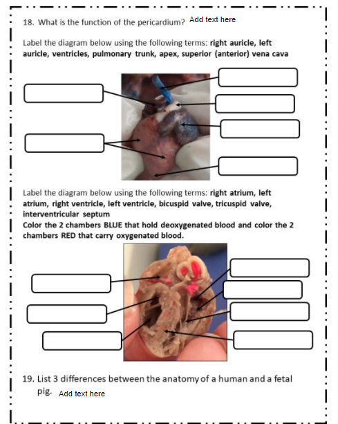
3. Insert your dissecting scissors or scalpel into the left auricle at the base. of the aorta and make an incision down through the wall of the left atrium and ventricle, as shown by the dotted line in the external heart picture. Locate the mitral valve (or bicuspid valve) between the left atrium and ventricle. This will have two flaps of ...
The main difference between atrium and auricle is that atrium is a compartment of the heart whereas auricle is a small out-pouching of the atrium. Heart is composed of two atria and two ventricles. Atria are the upper compartments while ventricles are the lower compartments. Left atrium and right atrium are the two types of atria.
The left atrium is one of the four chambers of the heart, located on the left posterior side. Its primary roles are to act as a holding chamber for blood returning from the lungs and to act as a ...
The upper chamber of the heart where blood is entered is referred to as auricle or atrium. The human heart possesses two atria, left atrium and right atrium. The two atria are separated into the left atrium and right atrium by a muscular wall at the medial edge of the right atrium. This is referred as an interatrial septum. This
CT and MRI measurements of the left atrium rely on echocardiography data 7. Measurement is made at end ventricular systole when the left atrium is the maximum size. normal left atrial AP diameter. women: <4.0 cm. men: <4.1 cm. An accurate AP measurement is made on the three-chamber view on a gated cardiac CT.
In the diagram, where is the left auricle of the left atrium? G. In the diagram, where is the ascending aorta? ... labeled structure shown in the diagram is a pouch-like extension that serves to slightly increase the capacity of an atrium? G. What labeled structure in the figure is the ligamentum arteriosum? D.
The left atrium's job is to collect this blood and pump it to the left ventricle, which in turn pumps the blood to the body's tissues. Where is the auricle of the heart? The upper chambers on each side of the septum are auricles, the lower chambers are called ventricles.
Left Auricle. The left auricle, also known as the left atrial appendage (LAA), is a flap of heart wall on the anterior surface of the left atrium of the heart. Often confused with the left atrium, it is one of the most prominent structural features of the left atrium and plays an important role in the pumping of blood within the heart.
Function of Auricle/Physiology. In comparison, atrium is much smaller then ventricle. So to fill the ventricle with maximum blood it should have more blood holding capacity which accounts for the presence of Auricle. During Cardiac Cycle of 0.8 seconds when the normal heartbeat is 72 beats per minute.
The left auricle, also known as the left atrial appendage (LAA), is actually a small, muscular pouch at the upper corner of the left atrium. It collects oxygenated blood as it leaves the lungs and...
In the diagram, where is the left auricle of the left atrium? asked Sep 25, 2015 in Anatomy & Physiology by Kristan. a) C b) F c) G d) H e) I. anatomy-and-physiology ... Before birth, blood bypasses from the right atrium to the left atrium through the foramen ovale. In the adult, what structure is the remnant of the foramen ovale? ...
Left atrial appendage. The left atrial appendage (LAA) (also known as left auricle ) is a pouch-like projection from the main body of the left atrium, which lies in the atrioventricular sulcus in close proximity to the left circumflex artery, the left phrenic nerve, and the left pulmonary veins.
In the diagram, where is the left auricle of left atrium? B. ... In the diagram, this supplies the walls of the left ventricle with oxygenated blood. ALL OF THE ABOVE (a, b, f, e) In the diagram, all of the following carry oxygenated blood. B. In the diagram, where is the marginal branch? F.
The left atrium has a distinctive appendage that is a finger-like pouch extending from the main body of the atrium. The main body then comprises of the pulmonary venous portion, the septal portion, and the vestibule, which is the outlet part of the atrial chamber surrounding the mitral orifice.
(a) Right auricle and right ventricle (b) Left auricle and left ventricle (c) Right auricle and left auricle (d) Right ventricle and left ventricle Answer: (a) Right auricle and right ventricle. Question 11. Rhythmic heartbeat is maintained by a highly specialized excitatory and conductive system. The correct sequence of events will be
Identify the letter that indicates the left auricle. D. Identify the letter that indicates the left common carotid artery. A. ... T/F-Oxygen rich blood returning from the lungs enters the left atrium through two right and two left pulmonary veins. True. T/F-The left ventricle walls are typically thicker than the right ventricular wall.
Disclaimer. All content on this website, including dictionary, thesaurus, literature, geography, and other reference data is for informational purposes only.
Transcribed image text: My Courses <Ex. 30: Best of Homework - Anatomy of the Heart Course Home Art-labeling Activity: Figure 30.2c (1 of 2) 5 of 12 > Syllabus Drag the labels onto the diagram to identify the parts and vessels of the heart (posterior view, 1 of 2). Scores Reset Help e Text Study Area Aorta Superior vena cava Document Sharing Left atrium User Settings Course Tools Auricle of ...
No, the left auricle is not the same as the left atrium. The auricles of the heart are portions of tissue that extend outward from the atria and connect to the blood vessels that deliver blood to ...
/heart_electrical_system-597907ca03f4020010e78125.jpg)

:background_color(FFFFFF):format(jpeg)/images/article/en/the-atria-of-the-heart/6ZerV5cTkzKIYq2s1n75Nw_Left_ventricle_and_atrium_of_hear.png)








/human-heart-circulatory-system-598167278-5c48d4d2c9e77c0001a577d4.jpg)
:watermark(/images/watermark_5000_10percent.png,0,0,0):watermark(/images/logo_url.png,-10,-10,0):format(jpeg)/images/overview_image/376/POnl8c3s8zUixaZCEChZ0g_left-atrium-and-ventricle_english.jpg)










0 Response to "42 in the diagram, where is the left auricle of the left atrium?"
Post a Comment