41 sheep brain labeled diagram
Sheep Brain Quiz - PurposeGames.com How is Your Memory? 11p Text Game. METRIC SYSTEM - the basics 10p Image Quiz. Countries of the European Union (by shape) 27p Image Quiz. 13 Colonies Quiz 13p Image Quiz. Sightseeing the US 28p Image Quiz. Math Theorems and Constants 14p Image Quiz. PG Lightning Game: Speed counting 20p Shape Quiz. Sheep brain dissection | Human Anatomy and Physiology Lab ... The sheep brain is quite similar to the human brain except for proportion. The sheep has a smaller cerebrum. Also, the sheep brain is oriented anterior to posterior (more horizontally), while the human brain is oriented superior to interior (more vertically.) Materials. Dissection tools and tray, lab gloves, preserved sheep brain.
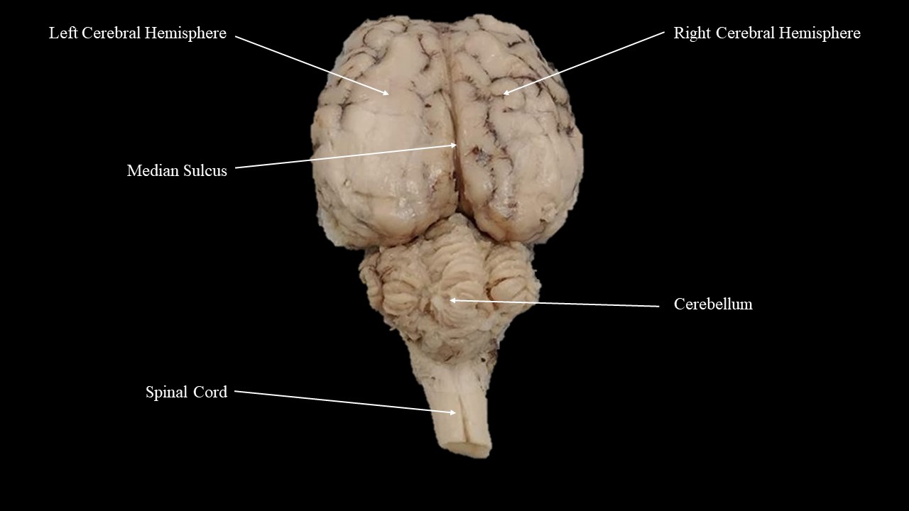
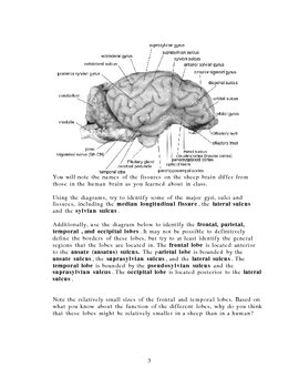
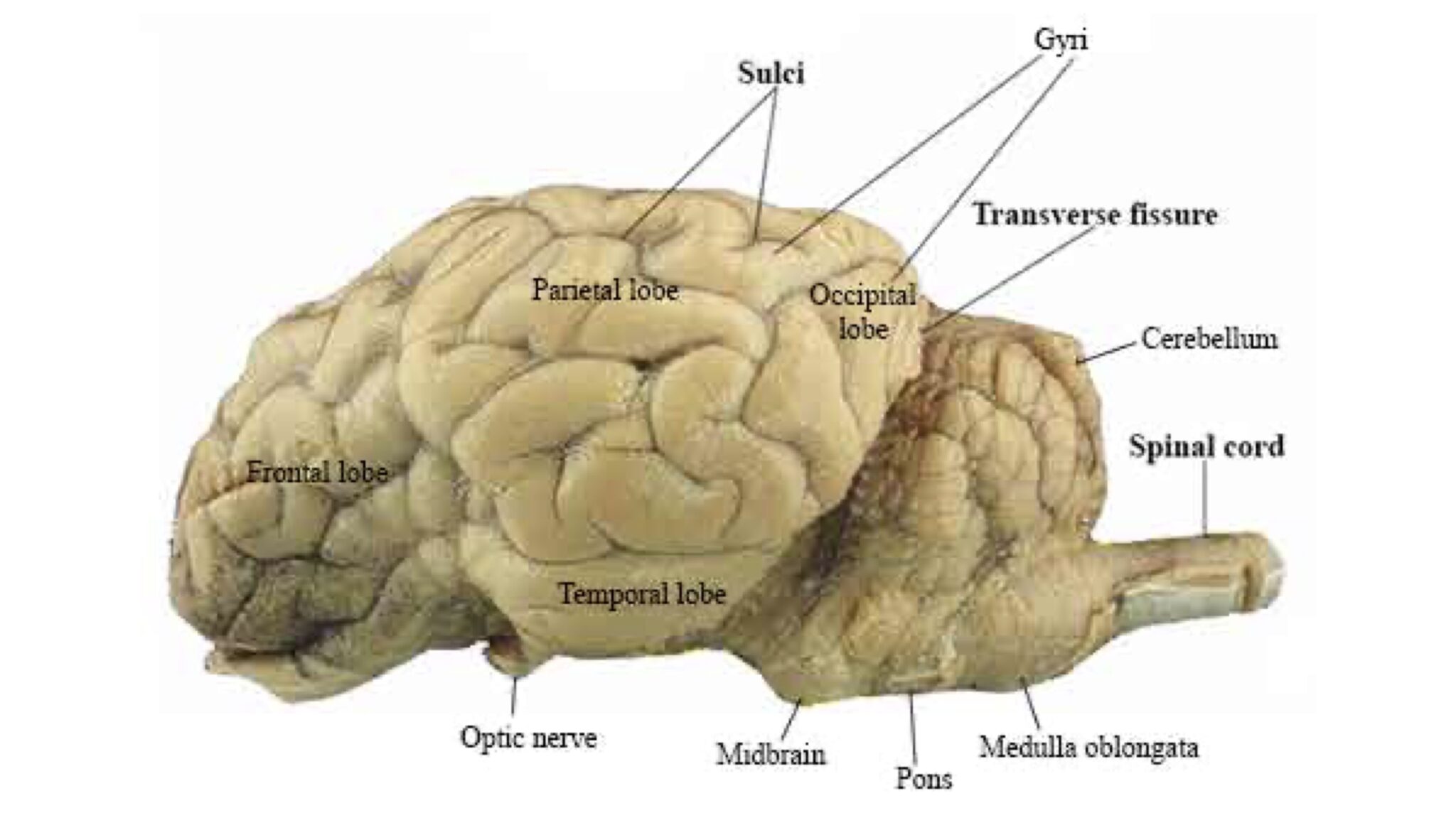
Sheep Brain Dissection Lab The lobes of the brain are visible, as well as the transverse fissure, which separates the cerebrum from the cerebellum. The convolutions of the brain are also visible as bumps (gyri) and grooves (sulci). Use the diagram below to help you locate these items. Dorsal View of the Sheep Brain . 8.
Sheep brain labeled diagram
Sheep Brain Labeled Diagram - Diagram Sketch Sheep Brain Labeled Diagram. angelo on January 2, 2022. Horse Brain 2 Brain Anatomy Brain Diagram Nervous System Anatomy. Labeled Diagram Of Brain Midsagittal View Diigo Groups Brain Anatomy Anatomy And Physiology Human Brain Anatomy. Sheep Brain Dissection Guide With Pictures Worksheets Nervous System Anatomy Brain Anatomy Dissection. Image ... DOC Sheep Brain Anatomy Lab Manual - amherst.edu Sheep Brain Anatomy Lab Manual. Based on original material by R. N. Leaton, Dartmouth College. Contributors to this version: Al Sorenson, Lisa Raskin, Sarah Turgeon, Steve George, and JP Baird. I. Introduction. The brain of the sheep is useful for study because its anatomy is similar to human brain anatomy. Although exact proportions (and names ... PDF Sheep Neuroanatomy Lab- Labeling Worksheet Psychology 2315 ... Sheep Neuroanatomy Lab- Labeling Worksheet Psychology 2315- Brain and Behaviour Kwantlen Polytechnic University Figure 1: Dorsal view Cerebellum, Frontal lobe, Occipital lobe, Parietal lobe, and Temporal lobe. Temporal Parietal Lobe Frontal Lobe Cerebellum Occipital Lobe
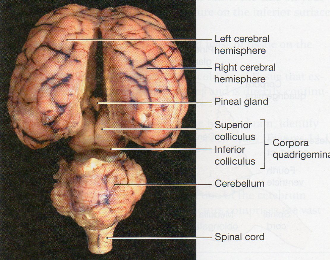
Sheep brain labeled diagram. Sheep Brain Anatomy Quiz - ProProfs Quiz Sheep Brain Anatomy Quiz. Sheep are wonderful and cute. The brain is an interesting organ. It helps with cognition and memory. Almost all the basic task In the body is commanded by the Brain. It is the control center of the body which regulates and control the process crucial for survival Are you interested in learning more about the brain of ... PDF DISSECTION OF THE SHEEP'S BRAIN - Hanover College DISSECTION OF THE SHEEP'S BRAIN Introduction The purpose of the sheep brain dissection is to familiarize you with the three-dimensional structure of the brain and teach you one of the great methods of studying the brain: looking at its structure. One of the great truths of studying biology is the saying that "anatomy precedes physiology". Sheep brain dissection directions F21.docx - Sheep Brain ... Sheep Brain Dissection Sheep brains, although much smaller than human brains, have similar features and can be a valuable addition to anatomy studies. See for yourself what the cerebrum, cerebellum, spinal cord, gray matter, white matter, and other parts of the brain look like! External Anatomy 1. You will be provided a preserved sheep brain for the dissection. Sheep Brain - Veterinary Anatomy Website Home Page The rostral colliculus(large arrow label) and the caudal colliculus(small arrow label) together form the tectumof the midbrain. Also labeled are the pineal body(green), the caudate nucleus(1), the floor of the fourth ventricle(white and pink) and cerebellar peduncles(blue = rostral, red = middle, and yellow = caudal). Go Top
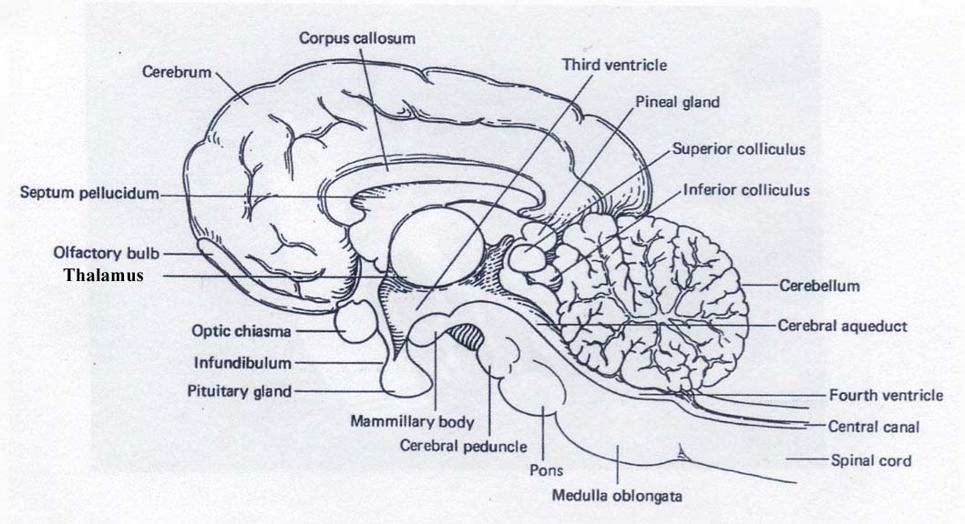
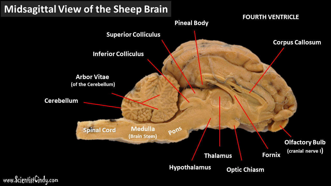
PDF Lab: Sheep Brain Dissection - Mrs. Moretz's Science Site to anatomy studies. See for yourself what the . cerebrum, cerebellum, spinal cord, gray matter, white matter, and other parts of the brain look like! Observation: External Anatomy . 1. You'll need a . preserved sheep brain. for the dissection. Set the brain down so the flatter side, with the white . spinal cord. at one end, rests on the ... PDF Sheep Brain Midsagittal Section - Dr. Scott Croes' Website Sheep Brain -Parasagittal Section 1. Gray Matter 2. White Matter 3. Corpus Callosum 4. Lateral Ventricle 5. Caudate Nucleus 6. Septum Pellucidum 7. Fornix 8. Optic Chiasma 9. Third Ventricle 10. Thalamus (Ovid Nuclear Mass of Thalamus) 11. Corona Radiata 12. Hippocampus 13. Cerebral Aqueduct (of Sylvius) 14. Pituitary Gland (hypophysis) 15. PDF Neuroanatomy: Dissection of the Sheep Brain Examine the sheep brain with the membranes intact. You should be able to identify and use the following directional terms: Anterior / Posteriorfront / back Rostral / Caudal towards the beak / towards the tail Medial / Lateral towards the middle / towards the side Dorsal / Ventral top / bottom (on the CNS of a quadruped) PDF Distance Learning Program Anatomy of the Human Brain/Sheep ... function, and pathology. Those students participating in Sheep Brain Dissections will have the opportunity to dissect and compare anatomical structures. At the end of this document, you will find anatomical diagrams, vocabulary review, and pre/post tests for your students. The following topics will be covered: 1.
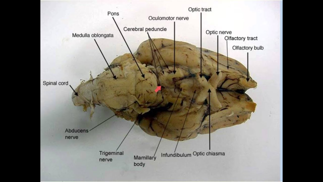
Sheep brain labeling Quiz - PurposeGames.com 25. 100% needed. Something different? METRIC SYSTEM - the basics 10p Image Quiz. Famous Hats (part 1) 10p Shape Quiz. Mountain Ranges of the World 44p Image Quiz. Continents 6p Image Quiz. # SUDOKU # 10p Image Quiz. Country Shapes - Slide Quiz 19p Image Quiz. Sheep Brain Label - The Biology Corner Label theBrain of the Sheep. Publisher: Biologycorner.com; follow on Google+ This work is licensed under a Creative Commons Attribution-NonCommercial 3.0 Unported License. Brain Label Answer Key. Image adapted from a photograph of the sheep brain. ... Diagram of Sheep Brain - Lateral view - Modesto Junior College Diagram of Sheep Brain - Lateral view - Modesto Junior College ALEX | Alabama Learning Exchange Students will use a Venn diagram to compare lightning and static electricity. Then, students will experiment with static electricity and read nonfiction passages about lightning and lightning rods. Finally, they will apply their learning to construct a model of a lightning rod system that protects a house from a lightning-induced fire.
Topic: Sheep Brain Dissection Grades: 8-12th Number of ...
Sheep Brain Dissection labeled Diagram | Quizlet Start studying Sheep Brain Dissection labeled. Learn vocabulary, terms, and more with flashcards, games, and other study tools.
sheep brain anatomy
Brain Lobes Diagram Labeled - Studying Diagrams With more related things such sheep brain diagram labeled brain nervous system worksheet and blank heart diagram. The diagram of the brain is useful for both Class 10 and 12. THE LOBES Occipital lobe Lower back of the brain. BI 335 Advanced Human Anatomy and Physiology Western Oregon University BRAIN ANATOMY Adapted from Human Anatomy ...
Physiological Psychology
Sheep Brain Dissection labeled 2 Diagram - Quizlet Start studying Sheep Brain Dissection labeled 2. Learn vocabulary, terms, and more with flashcards, games, and other study tools.
Physiological Psychology
A virtual sheep brain dissection guides anatomy studies ... Jun 6, 2018 - A virtual sheep brain dissection guides anatomy studies with photos & blank diagrams. Also shop complete dissection kits: guide, tools & preserved specimen.

BIO201-Sheep Brain
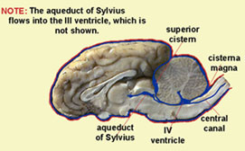
Functions - Sheep Brain Dissection Hypothalamus- is a portion of the brain that contains a number of small nuclei with a variety of functions. One of the most important functions of the hypothalamus is to link the nervous system to the endocrine system via the pituitary gland. Cerebral Acquesduct-a fluid-filled canal that runs through the midbrain connecting the third and fourth ...
Sheep Neuroanatomy Lab- Labeling Worksheet Figure 1: Dorsal view
Sheep Brain Anatomy and Function Flashcards - Cram.com Study Flashcards On Sheep Brain Anatomy and Function at Cram.com. Quickly memorize the terms, phrases and much more. Cram.com makes it easy to get the grade you want!

HSF Lab 1 - Exercise 17 (sheep brain) Diagram | Quizlet
Sheep Brain Label | Brain diagram, Dissection, Brain anatomy Sheep Brain Label. A drawing of the brain with the parts unlabeled. Students can practice naming the parts of the brain, then check their answers with the provided key. ... Download for free Brain Diagram #1647708, download othes brain anatomy for free. Sylvia Lopez. BRAIN. Human Skeleton Anatomy. Biology Poster. Vertebral Artery. Thoracic ...
11c Brain Anatomy
Sheep Brain Dissection with Labeled Images The sheep brain is exposed and each of the structures are labeled and described in a sequential manner, in the same way that a real dissection would occur. Sheep Brain Dissection. 1. The sheep brain is enclosed in a tough outer covering called the dura mater. You can still see some structures on the brain before you remove the dura mater.

Sheep Brain Diagram
Sheep Brain Diagram - Diagram Sketch Sheep Brain Diagram. angelo on October 9, 2021. Image Result For Sheep Brain Labeled Brain Diagram Human Brain Diagram Brain Anatomy. Sheep Brain Dissection Guide With Pictures Worksheets Nervous System Anatomy Brain Anatomy Dissection. Sheep Brain External View Labeled Anatomia Veterinaria Anatomia Veterinaria.
The Brain - SCIENTIST CINDY
S H E E P B R A I N L A B - Execulink To study the structure and function of the mammalian (sheep) brain. References: "Nelson Biology 12" (chapter 9), dissection charts, models, internet, other texts. Instructions - External Anatomy: Obtain a handout with diagrams of the brain from your teacher. Working in groups of two or three, select a sheep brain.
3.2: Sheep brain - Medicine LibreTexts
A new GABAergic somatostatin ... - Molecular Psychiatry 18-06-2020 · A negative control was performed with no injection of RG into the NAc shell, and no RV expressing dsRed labeled cells were found in the above brain regions (Fig. S5).

Sheep brain dissection | Human Anatomy and Physiology Lab ...
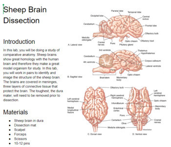
PDF Sheep Brain Dissection - Administration Sheep Brain Dissection The purpose of this exercise is to introduce you to the mammalian brain; you will use a sheep's brain. While the sheep brain differs from the human brain in many details, they both have the same basic anatomy, and, it is larger than the rat brain. Work in teams of four students.
Sheep Brain Dorsal View Ventral View Hippocampal ...
Sheep Brain Dissection Project Guide | HST Learning Center Sheep Brain Dissection: Internal Anatomy. Place the brain with the curved top side of the cerebrum facing up. Use a scalpel (or sharp, thin knife) to slice through the brain along the center line, starting at the cerebrum and going down through the cerebellum, spinal cord, medulla, and pons. Separate the two halves of the brain and lay them ...
Sheep Brain Dissection
PDF Sheep Neuroanatomy Lab- Labeling Worksheet Psychology 2315 ... Sheep Neuroanatomy Lab- Labeling Worksheet Psychology 2315- Brain and Behaviour Kwantlen Polytechnic University Figure 1: Dorsal view Cerebellum, Frontal lobe, Occipital lobe, Parietal lobe, and Temporal lobe. Temporal Parietal Lobe Frontal Lobe Cerebellum Occipital Lobe

Sheep brain - external anatomy | Sheep, Food, Brain
DOC Sheep Brain Anatomy Lab Manual - amherst.edu Sheep Brain Anatomy Lab Manual. Based on original material by R. N. Leaton, Dartmouth College. Contributors to this version: Al Sorenson, Lisa Raskin, Sarah Turgeon, Steve George, and JP Baird. I. Introduction. The brain of the sheep is useful for study because its anatomy is similar to human brain anatomy. Although exact proportions (and names ...

Lab - Sheep Brain Dissection
Sheep Brain Labeled Diagram - Diagram Sketch Sheep Brain Labeled Diagram. angelo on January 2, 2022. Horse Brain 2 Brain Anatomy Brain Diagram Nervous System Anatomy. Labeled Diagram Of Brain Midsagittal View Diigo Groups Brain Anatomy Anatomy And Physiology Human Brain Anatomy. Sheep Brain Dissection Guide With Pictures Worksheets Nervous System Anatomy Brain Anatomy Dissection. Image ...
Medical Detectives Lesson 27
Sheep Brain Dissection with Labeled Images
11.7: Sheep Brain Dissection - Biology LibreTexts
External sheep brain dissection guide
09.06.09: Brain Structure and Function and Disease
A&P 2 Lab page 7

Sheep Brain Dissection | Carolina.com

Dissecting Sheep Brains With Sixth Graders | Brains Explained
DISSECTION OF THE SHEEP'S BRAIN
Sheep Brain Dissection Teaching Resources | Teachers Pay Teachers

Sheep brain images | Lab | Amherst College

Sheep Brain Images

BIOL 160: Human Anatomy and Physiology
SCB209 - Lab2 - Natural Sciences Open Educational Resources

Resources for Teaching Mammalian Neuroanatomy Using Sheep ...

Sheep Brain Dissection – Krysta H, Rinda G, Keira N, Cindy M ...
Sheep Brain Dissection Report

Sheep Brain

Sheep Brain Dissection Lab Companion
Sheep Brain Dissection with Labeled Images
Lab2

Sheep brain - external anatomy (ventral) | Hypoglossal nerve ...
Lab: Sheep Brain Dissection
Sheep brain dissection worksheet
Lab: Sheep Brain Dissection
0 Response to "41 sheep brain labeled diagram"
Post a Comment