42 trigeminal nerve branches diagram
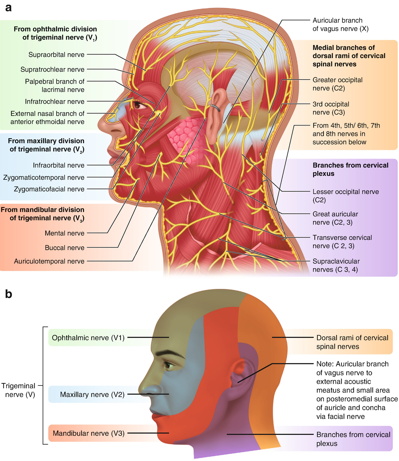
14 Jun 2021 — What are the trigeminal nerve branches? · Ophthalmic: This branch sends nerve impulses from the upper part of your face and scalp to your brain. "Trigeminal" = tri, and "-geminus" or twin, or thrice twinned derives from the fact that it has three major branches: Ophthalmic nerve (V1) 1st branch - sensory ...
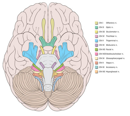
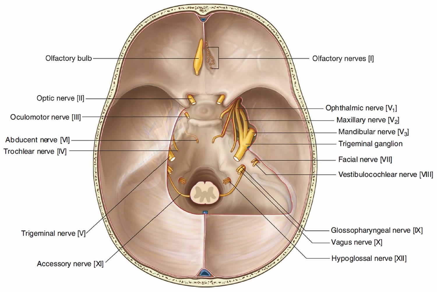
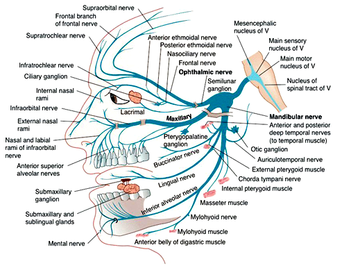
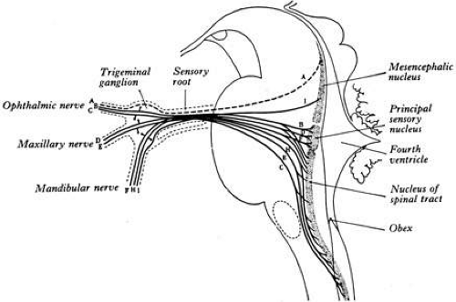
The trigeminal nerve, also known as the fifth (or V) cranial nerve, is a cranial nerve and its primary role is relaying sensory information from the face and head, although it does provide motor control to the muscles of mastication.It is both large and complicated and has multiple brainstem nuclei (sensory and motor) as well as many interconnections with other cranial nerves.
Trigeminal nerve branches diagram
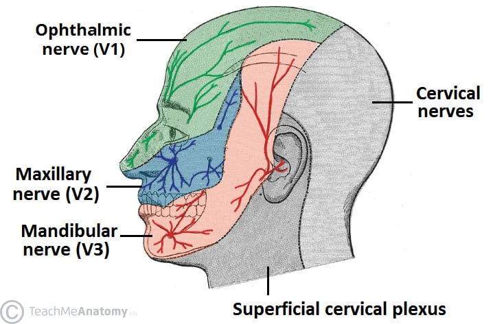
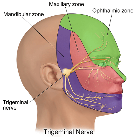
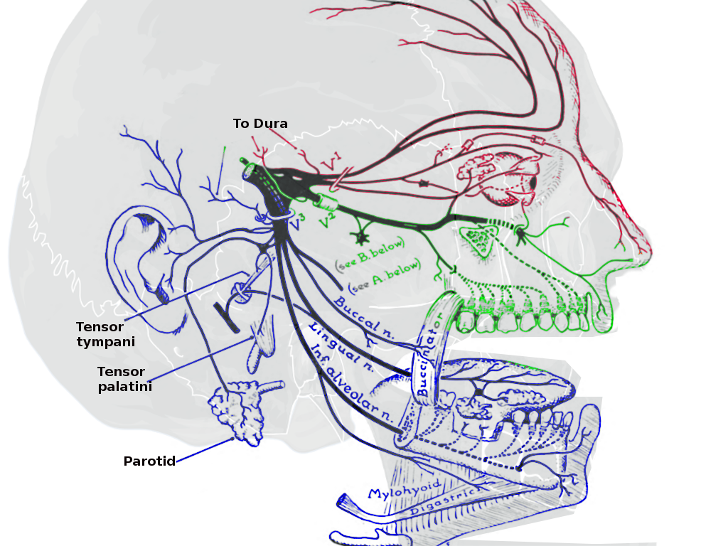
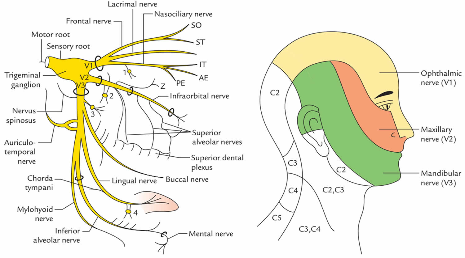
The trigeminal nerve is associated with derivatives of the 1st pharyngeal arch. Sensory: The three terminal branches of CN V innervate the skin, mucous membranes and sinuses of the face.Their distribution pattern is similar to the dermatome supply of spinal nerves (except there is little overlap in the supply of the divisions). Gross Anatomy. The trigeminal nerve is the largest and most complex of the 12 cranial nerves (CNs). It supplies sensations to the face, mucous membranes, and other structures of the head. It is the motor nerve for the muscles of mastication and contains proprioceptive fibers. The trigeminal nerve is the largest of the cranial nerves and can be further divided into three divisions: ophthalmic, maxillary, and mandibular. The ophthalmic division sends information from the scalp, forehead, and upper eye lids (the upper parts of the head) and is sensory in modality, of the general somatic sensory variety.
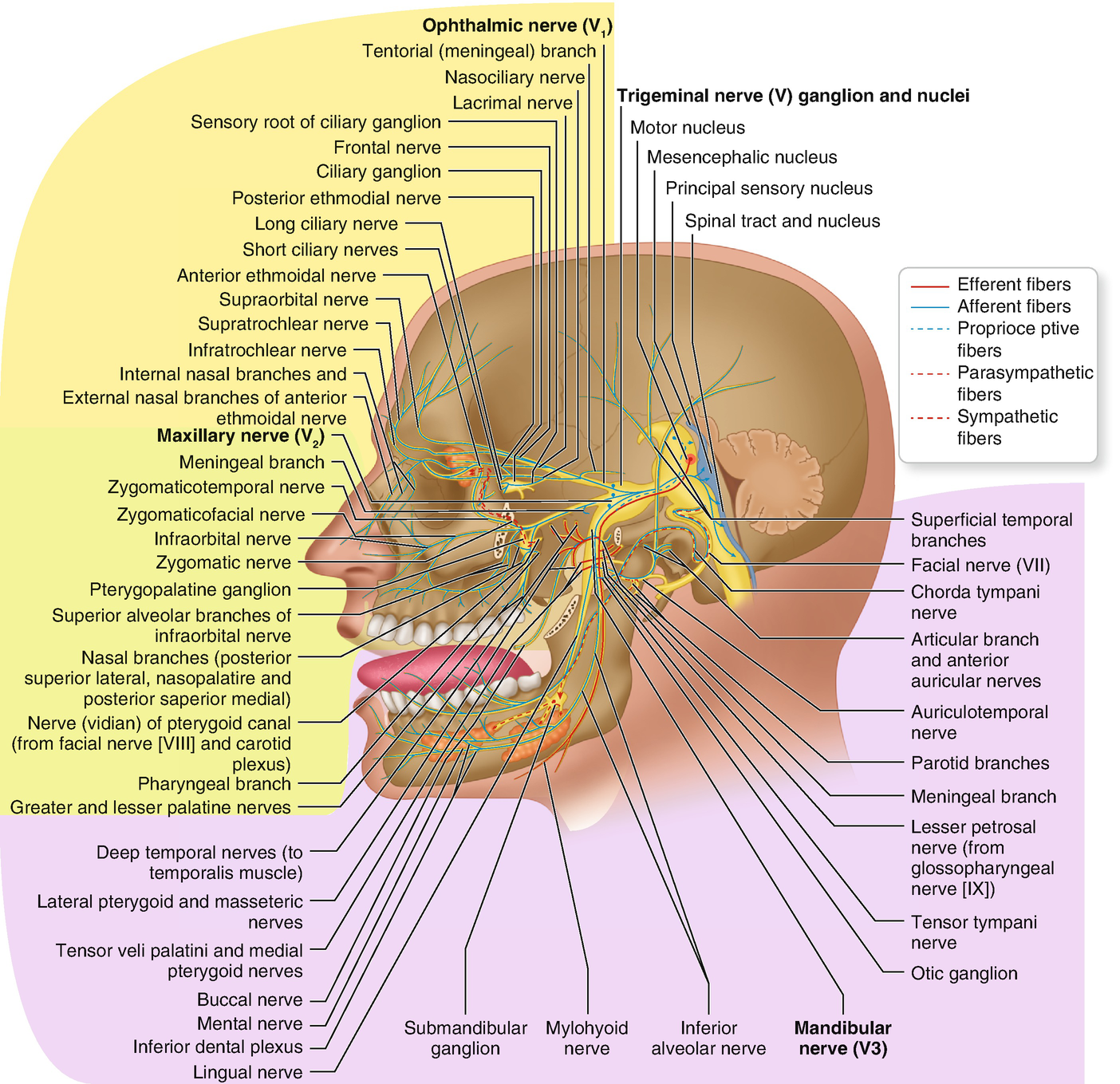
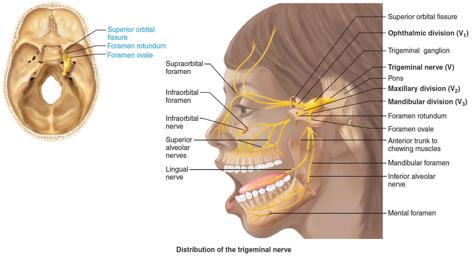
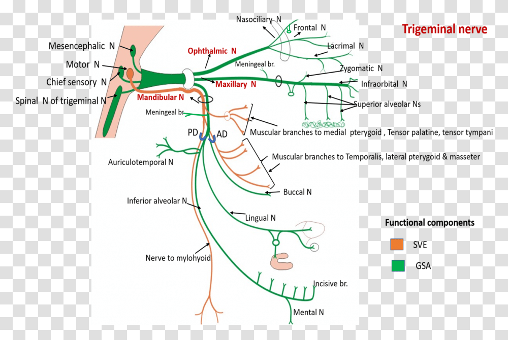
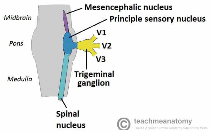
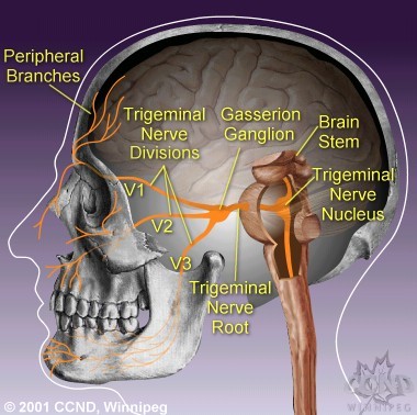
Trigeminal nerve branches diagram. The motor nerve branch of the trigeminal nerve is smaller than the sensory branches and exits from the brainstem through the root of the trigeminal nerve. Location . The trigeminal nerve roots and ganglion, like those of other cranial nerves, are located right outside the brainstem. The brainstem is the lower part of the brain that serves as ... The trigeminal nerve is the largest of the 12 cranial nerves. Its main function is transmitting sensory information to the skin, sinuses, and mucous membranes in the face. The three major branches of the trigeminal nerve—the ophthalmic nerve (V1), the maxillary nerve (V2) and the mandibular nerve (V3)—converge on the ...Sensory branches · Function · Trigeminal nucleus · Spinal trigeminal nucleus Branches of the trigeminal nerve. Print. Sections. Products and services. Trigeminal neuralgia results in pain occurring in an area of the face supplied by one or more of the three branches of the trigeminal nerve. There is a problem with information submitted for this request. Review/update the information highlighted below and resubmit the form.
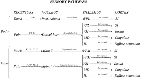
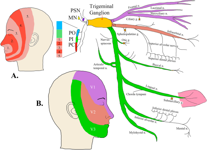
The trigeminal nerve is the fifth cranial nerve (CN V). Its primary function is to provide sensory and motor innervation to the face. The trigeminal nerve consists of three branches on either side that extend to different territories of the face. These branches join at the trigeminal ganglia which are located within the Meckel cave of the cranial cavity. The different branches are namely the ... Trigeminal nerve. The large trigeminal nerve or 5th cranial nerve has three branches: ophthalmic (V1), maxillary (V2), and mandibular (V3) divisions. Trigeminal nerve is a mixed nerve providing sensations of the face for touch, temperature, and pain from the upper, middle, and lower portions of the face, as well as the oral cavity, to the brain. The ophthalmic branch is the first division of the trigeminal nerve. It is a purely sensory nerve that carries afferent stimuli of pain, light touch, and temperature from the upper eyelids and supraorbital region of the face, up to the vertex of the head. The nerve also acts as a conduit for sympathetic fibers that require access to the ciliary body, lacrimal glands, cornea, and conjunctiva ... The trigeminal nerve is the largest of the cranial nerves and can be further divided into three divisions: ophthalmic, maxillary, and mandibular. The ophthalmic division sends information from the scalp, forehead, and upper eye lids (the upper parts of the head) and is sensory in modality, of the general somatic sensory variety.
Gross Anatomy. The trigeminal nerve is the largest and most complex of the 12 cranial nerves (CNs). It supplies sensations to the face, mucous membranes, and other structures of the head. It is the motor nerve for the muscles of mastication and contains proprioceptive fibers. The trigeminal nerve is associated with derivatives of the 1st pharyngeal arch. Sensory: The three terminal branches of CN V innervate the skin, mucous membranes and sinuses of the face.Their distribution pattern is similar to the dermatome supply of spinal nerves (except there is little overlap in the supply of the divisions).
Neural Blockade For Trigeminal Neuralgia Radiology Key

Trigeminal Nerve Wikipedia

Jual Terapi Akupunktur Untuk Nyeri Wajah Trigeminal Neuralgia Kab Magelang Klinik Pakualaman 2 Tokopedia

Anaesthesia Uk Anatomy Of The Trigeminal Nerve

Trigeminal Nerve Maxillary Branch Cn V2 Medical Discovery Tv
Trigeminal Nerve Radiology Reference Article Radiopaedia Org

Trigeminal Nerve Subdivisions Functional Components Structures Supplied Anatomy Qa
Trigeminal Nerve Anatomy Gross Anatomy Branches Of The Trigeminal Nerve Microscopic Anatomy
Anatomy Of The Trigeminal Nerve Springerlink
Trigeminal Nerve Cn V Anatomy Function And Branches Kenhub
Trigeminal Nerve Wikipedia

Anatomy Trigeminal Nerve
Trigeminal Nerve Anatomy Branches Distribution Function Damage Pain

The Trigeminal Nerve Branches Album On Imgur
The Trigeminal Nerve Cn V Course Divisions Teachmeanatomy
1
Trigeminal Nerve Anatomy Branches Distribution Function Damage Pain

Figure 2 From Microsurgical Reconstruction Of The Trigeminal Nerve Semantic Scholar

Maxillary Division Of The Trigeminal Nerve Gray S Illustration Radiology Case Radiopaedia Org
Trigeminal Nerve Course And Branches Of Trigeminal Nerve Plot Diagram Map Water Transparent Png Pngset Com

Anatomy Notes Map Of Trigeminal Update Another Re Scan J Flickr
The Trigeminal Nerve Cn V Course Divisions Teachmeanatomy

Integrative Neuroscience
Trigeminal Nerve The Definitive Guide Biology Dictionary
Facial And Trigeminal Nerve
The Fifth Cranial Nerve In Headaches The Journal Of Headache And Pain Full Text
Trigeminal Nerve Simplified Epomedicine

Trigeminal Nerve Illustration Radiology Case Radiopaedia Org

Anatomy Of The Trigeminal Nerve Sciencedirect
Trigeminal Nerve Branches

Our Responsibility The Trigeminal Nerve Oral Health Group

Trigeminal Nerve V And Its Brances Medical Illustration Medivisuals

Trigeminal Neuralgia Made Easy
How To Learn The Branches Of The Trigeminal Nerve With A Memory Palace Mullen Memory

The Trigeminal Nerve Human Anatomy
Your Complete Guide To Trigeminal Neuralgia A M Kaufmann M Patel Ccnd Winnipeg
Anatomy Of The Trigeminal Nerve Springerlink

Trigeminal Nerve Branches Radiology Students Of A M S Facebook
Neuroanatomy Cranial Nerve 5 Trigeminal Article
Trigeminal Nerve Anatomy Branches Distribution Function Damage Pain

Atlas Of Human Anatomy 1st Edition
Trigeminal Nerve Ento Key
0 Response to "42 trigeminal nerve branches diagram"
Post a Comment