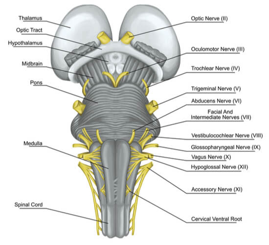
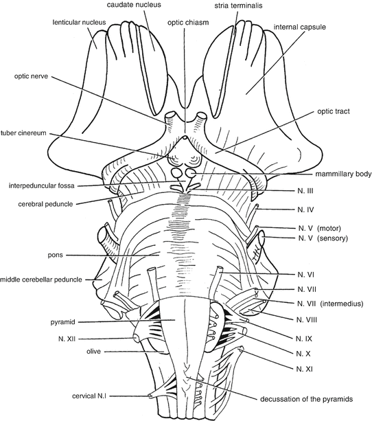
42 diagram of brain stem
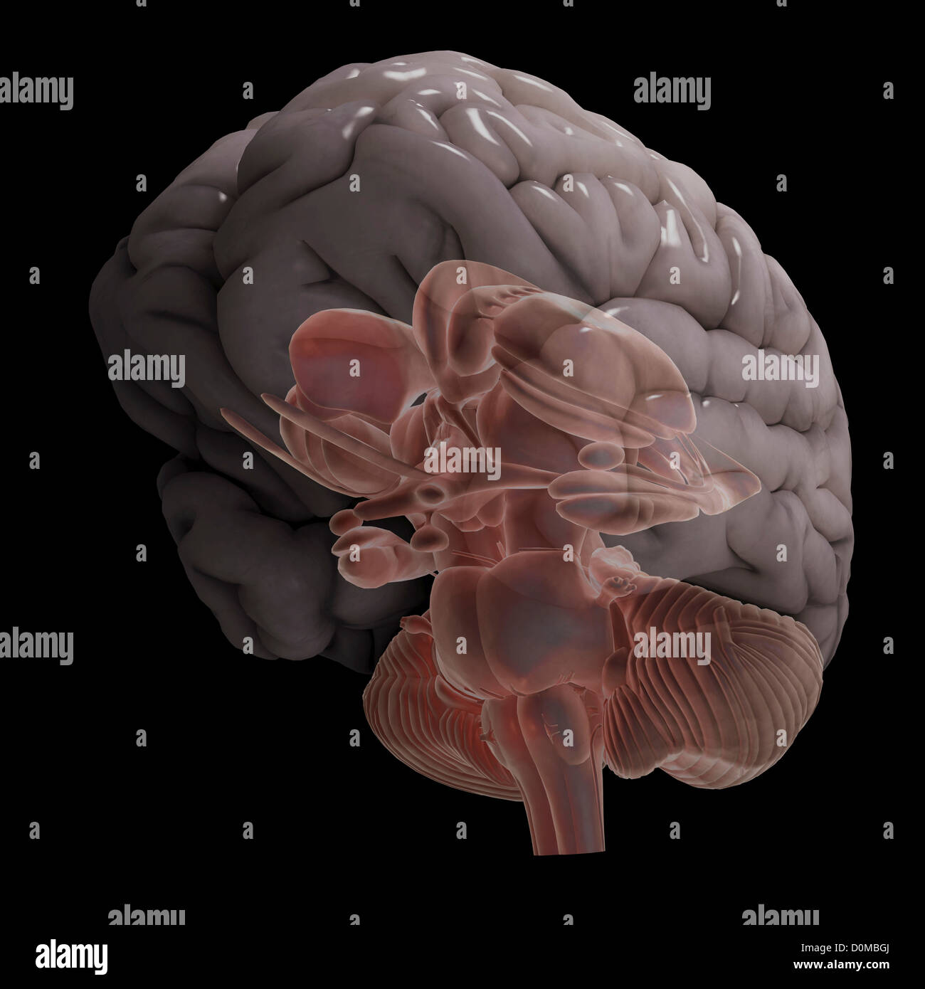
Brain Function Brainstem & Cerebellum Brainstem controls breathing, heartbeat, and articulate speech A stroke affecting the brain stem is potentially life threatening since this area of the brain controls functions such as breathing and instructing the heart to beat. Brain stem stroke may also cause double vision, nausea and loss of coordination. The brain stem also handles functions we usually do unconsciously, such as breathing, digesting food, heart activity, as well as maintains proper sleep cycles and body temperature. You can take this diagram of the brain quiz online to review the different parts of the brain and develop your understanding of its functions and structure.
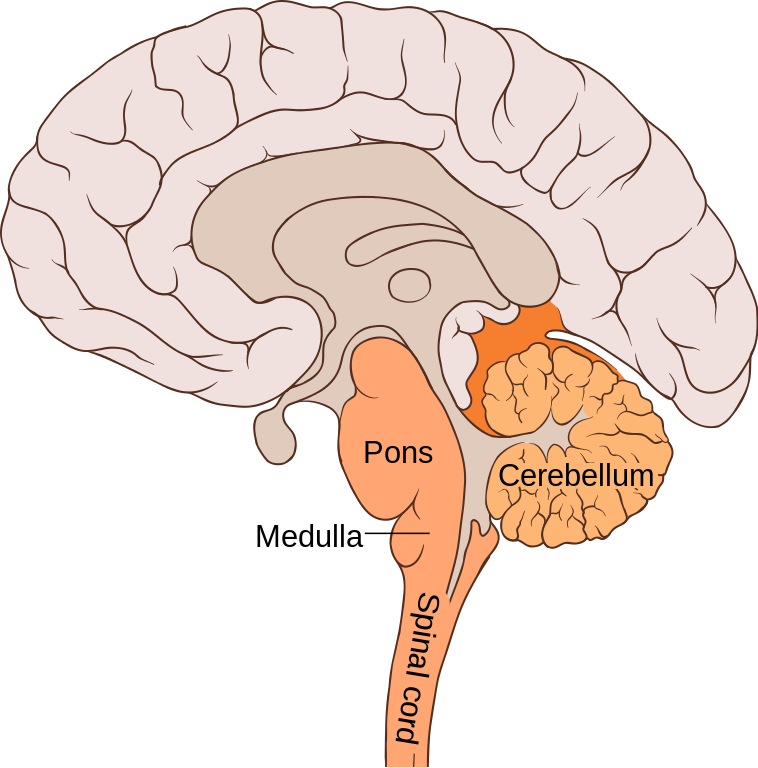
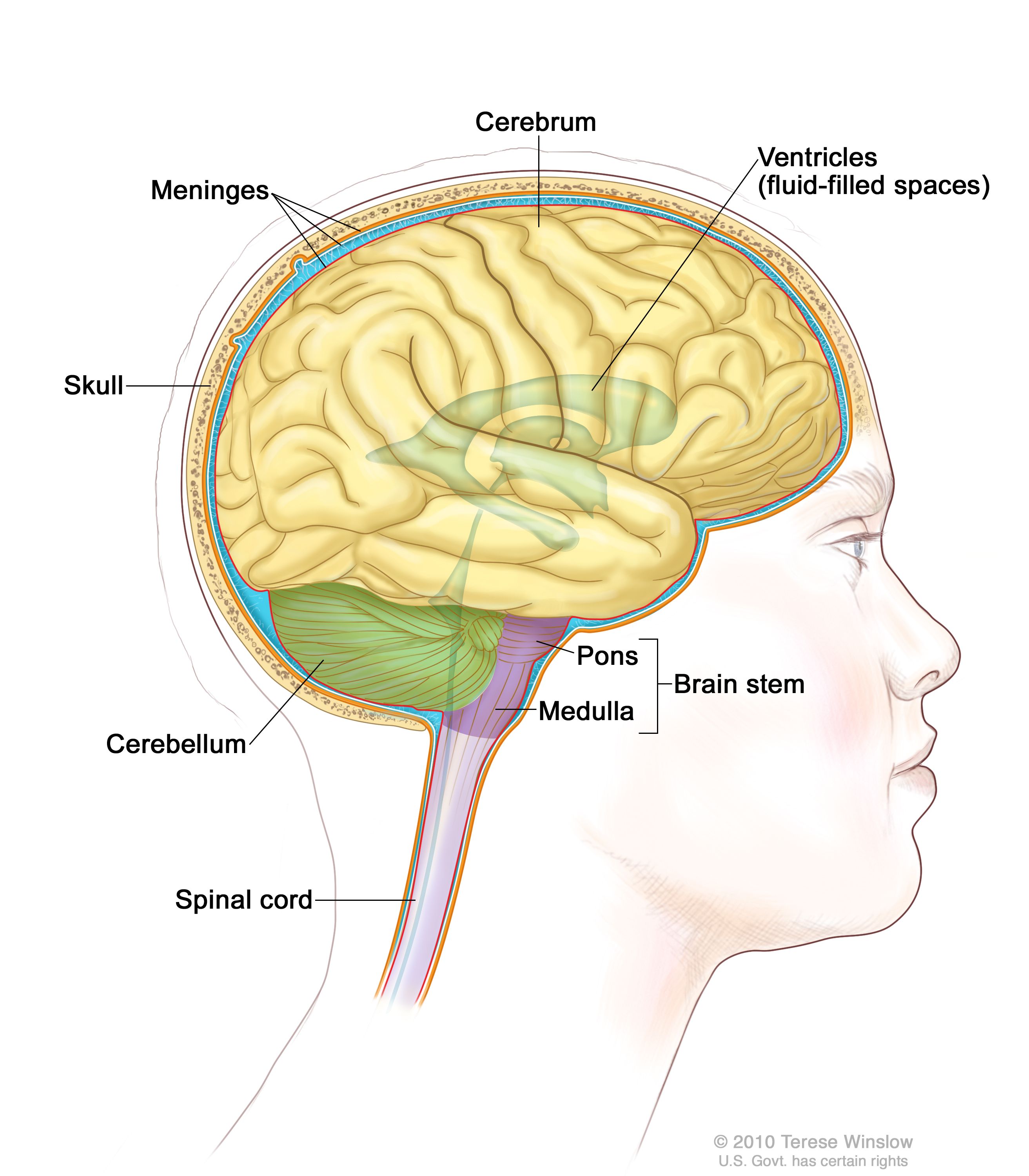
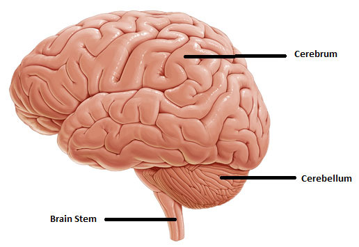
It is mainly composed of the Cerebellum, brain Stem, pons and the medulla. The cerebellum controls activities such as balance, coordination and muscle movements. The cerebellum is present at the backside of the brain at the base. The cerebellum is responsible for coordination and balance. This was a brief introduction to Diagram Of Brain.

Diagram of brain stem
Brain functions vector illustration. Labeled explanation organ parts scheme Brain functions vector illustration. Labeled explanation head organ parts scheme. Inner side view with educational section description. Cerebral cortex, hypothalamus, spinal cord and thalamus diagram. brain stem stock illustrations The midbrain (also called the mesencephalon) is the highest and first part of the brain stem.The midbrain consists of three general parts. The tectum (Latin: roof) is the highest part of the brain stem and forms the tissue that connects the brain stem to the lowest parts of the cerebellum. The main role of the tectum is to regulate reflex activity in response to visual and auditory stimuli and ... Brain Stem — The brain stem is located below the diencephalon and connects to the spinal cord, which if found inside the vertebrae of the spine. It can be divided into three structural parts that include the midbrain, pons, and medulla oblongata. (#8 in the diagram) 1. Midbrain: This area forms the upper part of the brain stem and functions ...
Diagram of brain stem. The Brainstem . Click On Any Label To Go To A Definition or Scroll Down To View All Definitions. Brainstem - The lower extension of the brain where it connects to the spinal cord.Neurological functions located in the brainstem include those necessary for survival (breathing, digestion, heart rate, blood pressure) and for arousal (being awake and alert). BRAIN STEM: The part of the brain that connects to the spinal cord. The brain stem controls functions basic to the survival of all animals, such as heart rate, breathing, digesting foods, and sleeping. It is the lowest, most primitive area of the human brain. CEREBELLUM: Two peach-size mounds of folded tissue located at the top of the brain ... This brain part controls thinking. This brain part controls balance, movement, and coordination. This brain part controls involuntary actions such as breathing, heartbeats, and digestion. This part of the nervous system moves messages between the brain and the body. This part of the cerebrum interprets and sorts information from the senses. The brainstem coordinates motor control signals sent from the brain to the body. This brain region also controls life-supporting autonomic functions of the peripheral nervous system.The fourth cerebral ventricle is located in the brainstem, posterior to the pons and medulla oblongata. This cerebrospinal fluid-filled ventricle is continuous with the cerebral aqueduct and the central canal of ...
The brainstem is the most caudal part of the brain.It adjoins, is structurally continuous with the spinal cord and consists of the:. midbrain (mesencephalon); pons (part of the metencephalon); medulla oblongata (myelencephalon); The brainstem provides the main motor and sensory innervation to the face and neck via the cranial nerves.It also provides the connection of the cerebrum, basal ... The brain, which is housed and protected by in the bones of the skull, makes up all parts of the central nervous system above the spinal cord. The brain can be divided into two major parts: the lower brain stem and the higher forebrain. The Brain Stem Boundless Anatomy And Physiology. Pathophysiology Of Hormonal Deficiency In Brain Death And Its Download Scientific Diagram. Pcbe Controversies In The Determination Of Death Chapter 3 The Clinical Presentation And Pathophysiology Of Total Brain Failure. Brain Stem Death Anaesthesia Intensive Care Medicine. Processes input from cerebral motor cortex, brain stem, and sensory receptors to provide precise coordinated movements of skeletal muscles via Purkinje cells (neurons of the cerebellum) Plays a major role in balance and posture
The brainstem (brain stem) is the distal part of the brain that is made up of the midbrain, pons, and medulla oblongata.Each of the three components has its own unique structure and function. Together, they help to regulate breathing, heart rate, blood pressure, and several other important functions.All of these brainstem functions are enabled because of its unique anatomy; since the brainstem ... Given below is a labeled diagram showing the brain stem and its related structures. Brain Stem and Structures. Cerebellum. The word 'cerebellum' literally means little brain. It is the second largest part of the brain, and is located at the back, below the occipital lobe, beneath the cerebrum and behind the brain stem. It contains an outer ... The brain stem begins inferior to the thalamus and runs approximately 7 cm before merging into the spinal cord. The brain stem centers produce the rigidly programmed, automatic behaviors necessary for survival. Positioned between the cerebrum and the spinal cord, the brain stem also provides a pathway for fiber tracts ... This interactive brain model is powered by the Wellcome Trust and developed by Matt Wimsatt and Jack Simpson; reviewed by John Morrison, Patrick Hof, and Edward Lein. Structure descriptions were written by Levi Gadye and Alexis Wnuk and Jane Roskams .
Human Brain Stem Diagram angelo. July 3, 2021. Diagram Of Cerebral Cortex Midbrain And Brainstem Brain Models Cerebral Cortex Medical Illustration . Pin On Erik Dalton . Biomed Illustrations Llc Offers Brain Anatomy Medical Illustration Exhibits For Jury Trials And Demand Package Brain Anatomy Brain Diagram Human Brain Diagram .
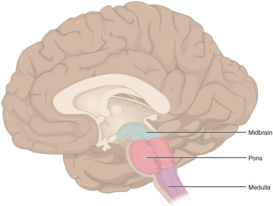
Parts of brainstem diagram. The brain stem is located in front of the cerebellum and connects to the spinal cord. The midbrain also called the mesencephalon is the highest and first part of the brain stemthe midbrain consists of three general parts. The brainstem is composed of the midbrain and portions of the hindbrain specifically the pons ...
Brain Stem — The brain stem is located below the diencephalon and connects to the spinal cord, which if found inside the vertebrae of the spine. It can be divided into three structural parts that include the mid-brain, pons, and medulla oblongata. (#8 in the diagram) 1. Midbrain: This area forms the upper part of the brain stem and functions ...
Brainstem. The brainstem, positioned at the base of the posterior region of the brain, functions as the critical point of connection between the central and peripheral nervous systems. There are twelve pairs of nerves that stem directly from the brainstem to control all senses that can be detected from the organs in the cranial region.
The study of the internal structure of the brain stem is shown with multiple diagrams in axial section showing the nuclei, tract, fibres and lemnisci. It should be noted that many structures are not represented for didactic purposes.
The brain stem connects the spinal cord to the higher-thinking centers of the brain. It consists of three structures: the medulla oblongata, the pons, and the midbrain. The medulla oblongata is continuous with the spinal cord and connects to the pons above. Both the medulla and the pons are considered part of the hindbrain.

Spinal Cord Label Diagram With Brain Stem Ppt Powerpoint Presentation Gallery Shapes Pdf Powerpoint Templates
Brain. The brain is one of the most complex and magnificent organs in the human body. Our brain gives us awareness of ourselves and of our environment, processing a constant stream of sensory data. It controls our muscle movements, the secretions of our glands, and even our breathing and internal temperature.
The brain stem contains the pons and medulla oblongata. Evolutionarily speaking, the hindbrain contains the oldest parts of the brain, which all vertebrates possess, though they may look different from species to species. Midbrain. The midbrain makes up part of the brain stem. It is located between the hindbrain and forebrain.
From the case: Brainstem cross-sectional anatomy (diagrams) Diagram. Entire brainstem. Upper midbrain. From the case: Brainstem cross-sectional anatomy (diagrams) Diagram. Upper midbrain. Lower midbrain. From the case: Brainstem cross-sectional anatomy (diagrams)
The brain stem is located in front of the cerebellum and connects to the spinal cord. It consists of three major parts: ... Use this interactive 3-D diagram to explore the brain. Brain conditions.
Location and Basic Physiology. In vertebrate anatomy, the brainstem is the most inferior portion of the brain, adjoining and structurally continuous with the brain and spinal cord. The brainstem gives rise to cranial nerves 3 through 12 and provides the main motor and sensory innervation to the face and neck via the cranial nerves.
Brain Stem — The brain stem is located below the diencephalon and connects to the spinal cord, which if found inside the vertebrae of the spine. It can be divided into three structural parts that include the midbrain, pons, and medulla oblongata. (#8 in the diagram) 1. Midbrain: This area forms the upper part of the brain stem and functions ...
The midbrain (also called the mesencephalon) is the highest and first part of the brain stem.The midbrain consists of three general parts. The tectum (Latin: roof) is the highest part of the brain stem and forms the tissue that connects the brain stem to the lowest parts of the cerebellum. The main role of the tectum is to regulate reflex activity in response to visual and auditory stimuli and ...
Brain functions vector illustration. Labeled explanation organ parts scheme Brain functions vector illustration. Labeled explanation head organ parts scheme. Inner side view with educational section description. Cerebral cortex, hypothalamus, spinal cord and thalamus diagram. brain stem stock illustrations






/bainstem-572261475f9b58857dc36e2e.jpg)
:background_color(FFFFFF):format(jpeg)/images/library/12362/brainstem-and-related-structures_english.jpg)


:background_color(FFFFFF):format(jpeg)/images/library/12358/anatomy-brainstem-anterior-view_english.jpg)




:max_bytes(150000):strip_icc()/GettyImages-1092334754-fd0644493b3148288970e38fd26aead0.jpg)
/profile-of-man-s-head-with-brain-anatomy-labeled-on-white-background-1093597090-f6a5470b98a4453997931b1cb72fb47d.jpg)



0 Response to "42 diagram of brain stem"
Post a Comment