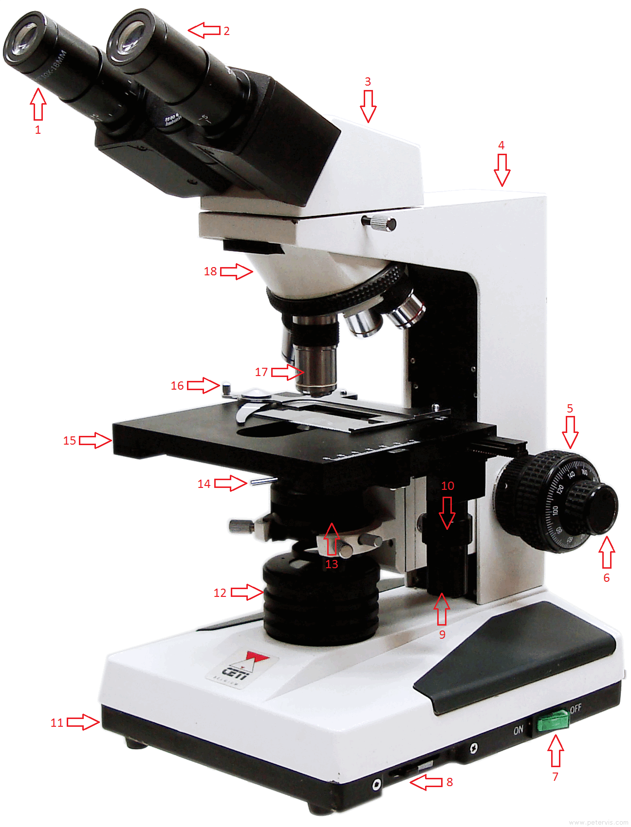
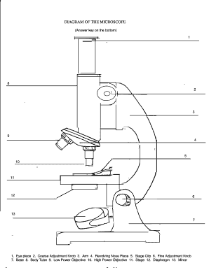
40 diagram of the microscope
750. The 10x objective of a compound microscope is being used to observe a specimen. If a total magnification of 150x is achieved, then the magnification of the ocular of this microscope must be. 15x. Base your answer to the following question on the diagram below and on your knowledge of biology. Essential Parts of Compound Microscope: (i) Lenses: (ii) Adjustment of Objective Lens: (iii) Stage: (iv) Mirror: ( ...
Revision Questions; Test your knowledge. Q. Define a Microscope. A. Microscopes are instruments that are used in science laboratories, to visualize very minute objects such as cells, microorganisms, giving a contrasting image, that is magnified. Q. State functions of a microscope. A. Microscope is usually used for the study of microscopic algae, fungi, and biological specimens.

Diagram of the microscope
Microscope Worksheet plant cell in a microscopic field of view is represented 10. Base your answer to the following question on the below. Microscope field of view 4000 urn The width (w) of this plant cell is closest to diagram below and on your knowledge of biology. The diagram shows cells as seen in the high-power (400> Fig. 1.1 is a labelled diagram of a leaf palisade mesophyll cell, as seen With a high quality light microscope. plasmodesma tonoplast vacuole cell wall Fig. 1.1 cytoplasm mitochondrion nucleus golgi body chloroplast An electron micrograph of the same leaf mesophyll cell at the same magnification would show more detail than is shown in Fig. 1.1. Microscope Parts and Functions With Labeled Diagram and Functions How does a Compound Microscope Work?. Before exploring microscope parts and functions, you should probably understand that the compound light microscope is more complicated than just a microscope with more than one lens.. First, the purpose of a microscope is to magnify a small object or to magnify the fine details of a larger ...
Diagram of the microscope. 1 Jun 2021 — The Microscopes parts divided into three different structural parts Head, Base, and Arms. Eyepiece Lens: the lens at the top that you look ... Print a microscope diagram, microscope worksheet, or practice microscope quiz in order to learn all the parts of a microscope. Microscope Parts and Functions Invented by a Dutch spectacle maker in the late 16th century, light microscopes use lenses and light to magnify images. Although a magnifying glass technically qualifies as a simple light microscope, today’s high-power—or compound— microscopes use two sets of lenses to give users a much In this video, you can learn how to draw diagram of microscope. In this video, you can learn how to draw diagram of microscope.
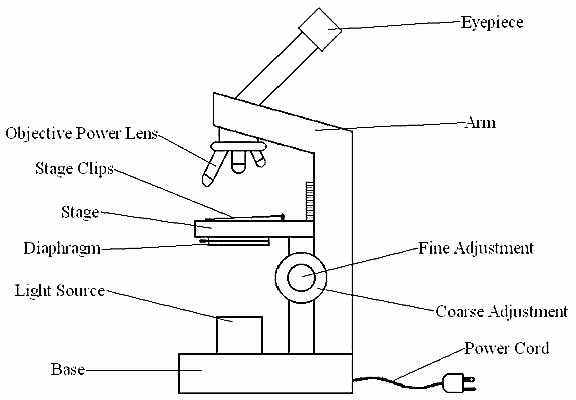
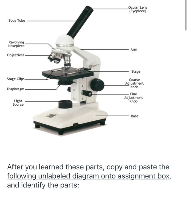
Microscope Labeled Diagram. Revision Questions : FAQ. How to tell the difference between a standard condenser and an Abbe condenser? Using a condenser, the illuminator’s light can be collected and focused upon a specimen. Under the microscope stage, near the diaphragm, they can be found. They are critical in obtaining crisp, clear images at ... or light microscope (vs./ an electron microscope) . The simplest optical microscope is the magnifying glass and is good to about ten times (10X) magnification. The compound microscope has two systems of lenses for greater magnification, 1) the ocular, or eyepiece lens that Nov 14, 2018 - A collection of microscope diagrams and worksheets for science class. Download them all in one convenient PDF, and select the version that's ... Q.5. What are the two main types of microscopes? Ans: Simple single-lens microscopes and compound or double lens microscopes are the two main types of light microscopes. Q.6. What are the \(14\) parts of a microscope? Ans: The following image shows all the parts of a compound microscope.
Microscope Parts and Functions With Labeled Diagram and Functions How does a Compound Microscope Work?. Before exploring microscope parts and functions, you should probably understand that the compound light microscope is more complicated than just a microscope with more than one lens.. First, the purpose of a microscope is to magnify a small object or to magnify the fine details of a larger ... Fig. 1.1 is a labelled diagram of a leaf palisade mesophyll cell, as seen With a high quality light microscope. plasmodesma tonoplast vacuole cell wall Fig. 1.1 cytoplasm mitochondrion nucleus golgi body chloroplast An electron micrograph of the same leaf mesophyll cell at the same magnification would show more detail than is shown in Fig. 1.1. Microscope Worksheet plant cell in a microscopic field of view is represented 10. Base your answer to the following question on the below. Microscope field of view 4000 urn The width (w) of this plant cell is closest to diagram below and on your knowledge of biology. The diagram shows cells as seen in the high-power (400>
The Microscope
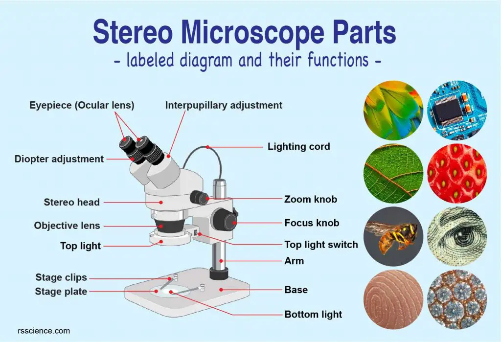
Parts Of Stereo Microscope Dissecting Microscope Labeled Diagram Functions And How To Use It
Microscope Safety And Diagram
Compound Microscope Types Parts Diagram Functions And Uses Laboratoryinfo Com
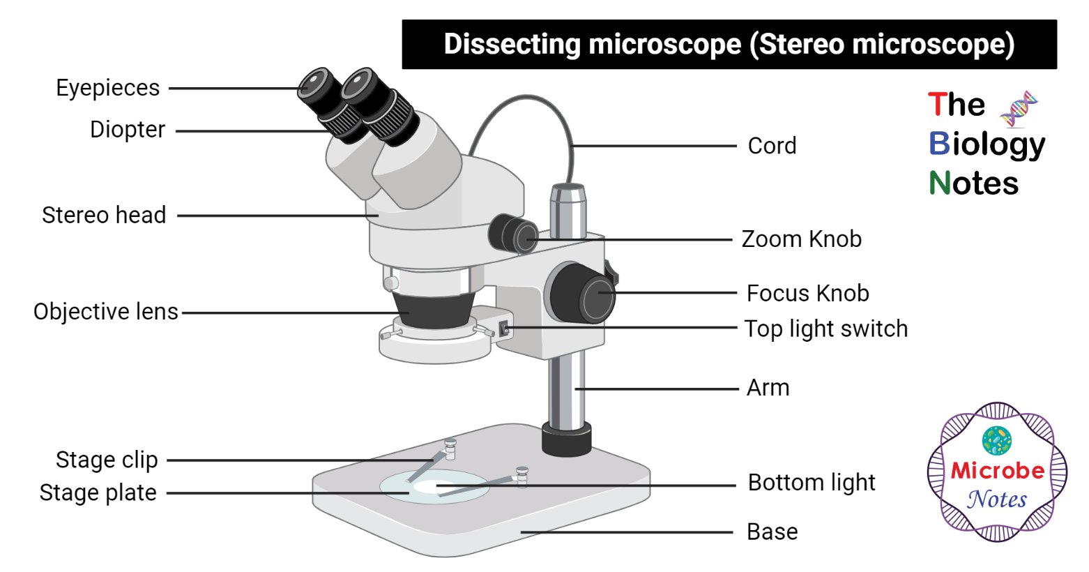
Dissecting Microscope Stereo Or Stereoscopic Microscope

Science Fair Project Ideas For Kids With Microscopes Microscope Parts Science Fair Projects Science Fair

White And Black Microscope Optical Microscope Light Scanning Electron Microscope Diagram Microscope Angle Technic Laboratory Png Pngwing

Microscope Diagram With Name Edusip

Parts Of A Microscope With Functions And Labeled Diagram
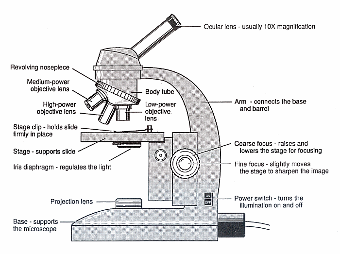
Diagram Of A Microscope Guide To Using A Microscope

The Compound Microscope Diagram Quizlet
30 Picture Of Microscope With Label Labels Database 2020

Compound Microscope Definition Parts Application Working Principle
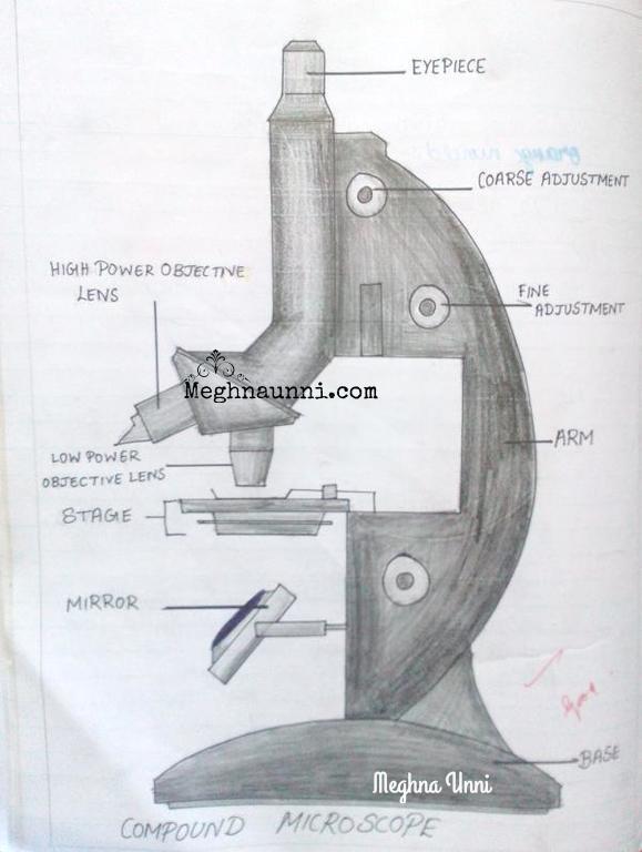
Biology Compound Microscope Diagram For Class 8 Meghnaunni Com
Solved Diagram Shows A Typical Light Microscope With Its Chegg Com

Schematic Diagram Of The Polarizing Microscope Download Scientific Diagram
1

Microscope Diagram Labeled Unlabeled And Blank Parts Of A Microscope

Schematic Diagram Of The Of The Wflpcf Microscope Download Scientific Diagram

How To Draw Compound Of Microscope Easily Step By Step Youtube

Compound Microscope Definition Labeled Diagram Parts Uses
Labelled Diagram Of Microscope Parts

20 Inspiration Draw Compound Microscope Perangkat Sekolah

Microscope Microscope Parts Labeled Diagram And Functions
Microscope Diagram Fill Online Printable Fillable Blank Pdffiller

Diagram Of A Compound Microscope
School Microscope Icon Outline Style Royalty Free Vector
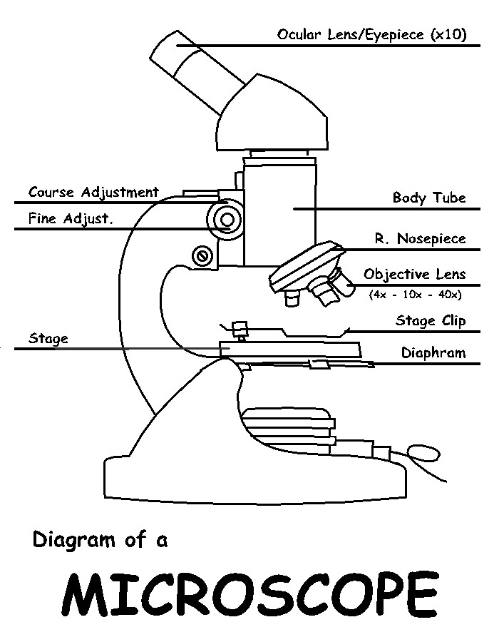
Diagram Clip Art Library
16 Parts Of A Compound Microscope Diagrams And Video Microscope Clarity
Monocular Light Microscope Labelled Diagram

Labeling The Parts Of The Microscope Microscope World Resources

Introduction Microscope Diagram Diagram Quizlet

Draw Microscope Diagram Sketch Template Diagram Coloring Pages Draw
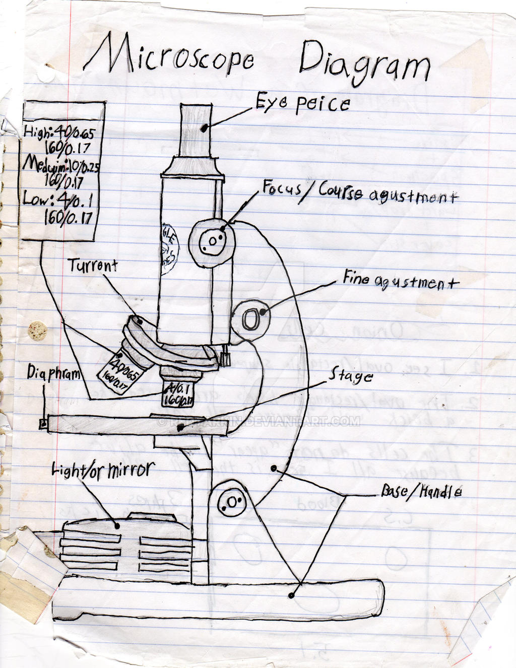
Microscope Diagram By Redtarkin On Deviantart

Parts Of Compound Microscope With Functions And Labelled Diagram

Carl Zeiss Microscopy Optical Microscope Worksheet Diagram Microscope Angle Technic Cell Png Pngwing

File Microscope Compound Diagram Png Wikimedia Commons

Microscope Drawing With Label Clip Art Library

Microscope Diagram Biology Printer Friendly Page Preproom Org

Label Microscope Diagram Charts Microscope Chemistry Lab Equipment Diagram Chart
0 Response to "40 diagram of the microscope"
Post a Comment